
- Submit a Protocol
- Receive Our Alerts
- EN

- Protocols

- Articles and Issues
- For Authors
- About
- Become a Reviewer

In vitro Cleavage and Electrophoretic Mobility Shift Assays for Very Fast CRISPR
Published: Vol 11, Iss 17, Sep 5, 2021 DOI: 10.21769/BioProtoc.4138 Views: 3009
Reviewed by: Pilar Villacampa AlcubierreWenyang Li

Original research article
The authors used this protocol in:
Jun 2020

Protocol Collections
Comprehensive collections of detailed, peer-reviewed protocols focusing on specific topics


Related protocols
Abstract
CRISPR-Cas9 has transformed biomedical research and medicine through convenient and targeted manipulation of DNA. Time- and spatially-resolved control over Cas9 activity through the recently developed very fast CRISPR (vfCRISPR) system have facilitated comprehensive studies of DNA damage and repair. Understanding the fundamental principles of Cas9 binding and cleavage behavior is essential before the widespread use of these systems and can be readily accomplished in vitro through both cleavage and electrophoretic mobility shift assays (EMSA). The protocol for in vitro cleavage consists of Cas9 with guide RNA (gRNA) ribonucleoprotein (RNP) formation, followed by incubation with target DNA. For EMSA, this reaction is directly loaded onto an agarose gel for visualization of the target DNA band that is shifted due to binding by the Cas9 RNP. To assay for cleavage, Proteinase K is added to degrade the RNP, allowing target DNA (cleaved and/or uncleaved) to migrate consistently with its molecular weight. Heating at 95°C rapidly inactivates the RNP on demand, allowing time-resolved measurements of Cas9 cleavage kinetics. This protocol facilitates the characterization of the light-activation mechanism of photocaged vfCRISPR gRNA.
Keywords: CRISPR-Cas9Background
Endonucleases, especially Cas9, from clustered regularly interspaced short palindromic repeats (CRISPR) and CRISPR-associated (CRISPR-Cas) adaptive immune systems (Marraffini et al., 2015) have found widespread use as highly versatile tools in the biomedical sciences from genome editing, functional screening, to imaging (Adli et al., 2018). Recently, highly resolved spatiotemporal control over Cas9 activity via very fast CRISPR (vfCRISPR) has enabled detailed studies of the DNA damage response through highly synchronized activation of Cas9 in space and time (Liu et al., 2020). For all uses, characterization of these nucleases is essential and can be most readily accomplished in vitro (Singh et al., 2016). Electrophoretic mobility shift assays (EMSA) are powerful tools to characterize protein-DNA binding that can be directly applied to determine the binding affinity of Cas9 to nucleic acids (Hellman et al., 2007). In contrast, in vitro cleavage assays allow characterization of the Cas9 DNA cleavage mechanism. In this manuscript, we outline detailed protocols for both strategies to characterize the properties of the vfCRISPR light-inducible Cas9 system. We demonstrate how both EMSA and in vitro cleavage assays characterize the binding, speed, and efficiency of vfCRISPR, which paves the way for its use in biological systems.
Materials and Reagents
10 mg/ml SpCas9 (purified in-house from BL21 CodonPlus (DE3)-RIL cells [Agilent 230245], but can also be purchased from Integrated DNA Technologies) (Alt-R®, catalog number: 1081058, storage temperature: -80°C)
tracrRNA (Integrated DNA Technologies) (Alt-R®, catalog number: 1072533, storage temperature: -80°C)
Photocaged crRNA (cgRNA) from vfCRISPR targeting PPP1R2 (BioSynthesis, sequence: GACUUCCUCUAUGGUGGCGUGUUUUAGAGCUAUGCUGUUUUG; U corresponds to replacements of uracil with NPOM-dT photocaged nucleotides, storage temperature: -80°C)
HEK293T cells (ATCC, catalog number: CRL-3216, storage temperature: liquid nitrogen vapor)
Nuclease-Free Duplex Buffer (Integrated DNA Technologies, catalog number: 11-01-03-01, storage temperature: 4°C)
PPP1R2 Fwd PCR primer (Integrated DNA Technologies, storage temperature: -20°C, sequence: 5’-GTTTCCGAGGCAGCAGTTG-3’)
PPP1R2 Rev PCR primer (Integrated DNA Technologies, storage temperature: -20°C, sequence: 5’-GCATGATAAACGTCATCGCCC-3’)
DNeasy Blood & Tissue Kit (Qiagen, DNeasy, catalog number: 69504)
QIAquick PCR purification kit (Qiagen, QIAquick, catalog number: 28104)
Q5® High-Fidelity 2X Master Mix (New England BioLabs, Q5®, catalog number: M0492, storage temperature: -20°C)
NEBufferTM 3.1 (New England BioLabs, catalog number: B7203S, storage temperature: -20°C)
Proteinase K, Molecular Biology Grade (New England BioLabs, catalog number: P8107S, storage temperature: -20°C)
E-GelTM Agarose Gels with SYBRTM Safe DNA Gel Stain, 4% (Thermo Fisher, E-GelTM, catalog number: A45206, storage temperature: room temperature)
100 bp DNA ladder (New England BioLabs, catalog number: N3231S)
HEPES (Sigma-Aldrich, catalog number: H3375)
KCl (Sigma-Aldrich, catalog number: P3911)
Glycerol (Sigma-Aldrich, catalog number: G5516)
NaOH (Sigma-Aldrich, catalog number: 221465)
Aluminum foil (Reynolds Wrap)
Nuclease-Free Water (not DEPC-treated) (InvitrogenTM, catalog number: AM9932)
Equipment
C1000 TouchTM Thermo Cycler (Bio-Rad, model number: 1851148), or any alternative thermocycler that can perform polymerase chain reaction (PCR)
E-Gel® iBaseTM Power System (Thermo Fisher, E-GelTM, catalog number: G6465), or alternative gel electrophoresis system
JAXMAN 365 nm LED flashlight (Amazon, JAXMAN, https://www.amazon.com/JAXMAN-Ultraviolet-365nm-Detector-Flashlight/dp/B06XW7S1CS/)
TyphoonTM FLA 9000 (GE Healthcare), or any alternative gel imager that can image ethidium bromide or SYBR Gold agarose gels
Procedure
Generation of target DNA at PPP1R2 for in vitro cleavage or EMSA
Purify genomic DNA (gDNA) from HEK293T cells using the DNeasy Blood & Tissue Kit following manufacturer’s instructions. Elute in 200 μl AE provided by the kit. Store gDNA in -20°C.
Prepare PCR primer mixture by mixing 10 μM of Fwd primer with 10 μM of Rev primer, both diluted in water.
Set up PCR reaction.
Component Volume Nuclease-Free Water (NFW) 3 μl Genomic DNA (10-50 ng) 1 μl Fwd/Rev primer mixture (10 μM) 1 μl Q5® High-Fidelity 2× Master Mix 5 μl Total 10 μl Start thermocycling protocol on the thermocycler.
Step Temp Time Initial Denaturation 98°C 30 s 35 cycles 98°C 10 s 68°C (PPP1R2) 10 s 72°C 20 s Final extension 72°C 2 min Hold 4°C Inf Extract PCR-amplified target DNA with the QIAquick PCR Purification Kit following manufacturer instructions. Elute in 30 μl EB provided by the kit.
Prepare 10 ml of Cas9 Dilution Buffer
Make solution composed of 20 mM HEPES, 500 mM KCl, and 20% glycerol.
Add sufficient 1 M NaOH to bring pH to 7.5.
Prepare 10 μM Cas9
Dilute 5 μl of 10 mg/ml Cas9 with 25 μl of Cas9 Dilution Buffer to make 30 μl of 10 μM SpCas9.
Anneal light-activatable crRNA with tracrRNA to form cgRNA
Resuspend photocaged crRNA and tracrRNA separately to 100 μM with Duplex Buffer.
Mix 3 μl of 100 μM photocaged crRNA with 3 μl of 100 μM tracrRNA in PCR tube.
Heat at 95°C for 3 min in the thermocycler with heated lid.
Cool on benchtop for 5 min.
Mix in 24 μl of Duplex Buffer to make 30 μl of 10 μM annealed cr/trRNA.
In vitro cleavage to test light activation capability of cgRNA
Mix the following components together in order. Thoroughly mix NFW and NEBufferTM 3.1 before adding cgRNA and Cas9 and mix again. Prepare 11 identical volumes, all in separate PCR tubes.
Component Volume NFW 8.1 μl NEBufferTM 3.1 1 μl 10 μM cgRNA targeting PPP1R2 0.5 μl 10 μM Cas9 0.4 μl Total 10 μl Leave on benchtop (room temperature) for 30 min to form RNP complex. Cover with aluminum film because the photocaged gRNA are light-sensitive.
Add 2 μl (~60 fmol) of PCR-amplified PPP1R2 target DNA to each RNP tube (11 in total). Mix well with pipette. For a highly efficient reaction, Cas9/cgRNA should be in at least 100-fold excess relative to target DNA. The target DNA should also be easily visible on an agarose gel with a gel imager.
Set up two thermocyclers – one to 37°C and the other to 95°C.
Move one tube to the 37°C thermocycler, open lid, and wait for 1 min. Turn on 365 nm LED flashlight 10 cm above tube for 1 s; then, immediately transfer tube to 95°C for 5 min and then to ice (which corresponds to ‘1 s’ sample) (Figure 1).
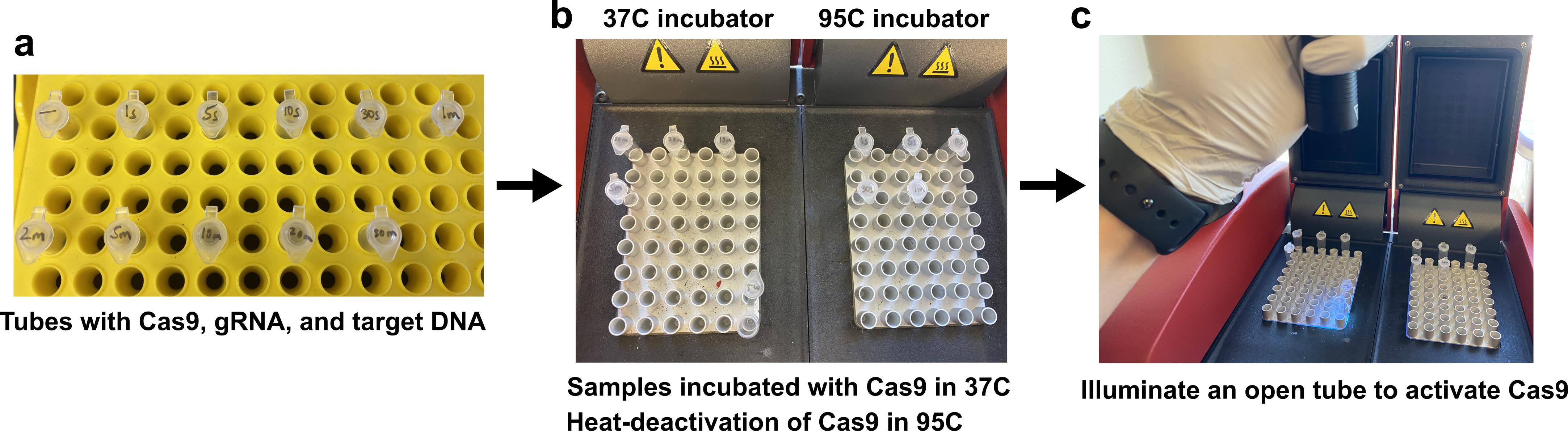
Figure 1. In vitro cleavage setup. (a) Cas9, gRNA, and target DNA are added to tubes labeled with the appropriate incubation times. (b) Tubes are first placed in the 37°C incubator. After light exposure and 37°C incubation for the specified time, each tube is transferred to the 95°C incubator for heat-deactivation of Cas9. (c) Light exposure using a commercially available LED flashlight. The tube is first opened, the flashlight is held 10 cm above the tube, and the light is turned on for 30 s.Repeat using three other tubes for 5 s, 10 s, and 30 s of light illumination (which corresponds to ‘5 s’, ’10 s’, and ’30 s’ samples, respectively).
Move another tube to the 37°C thermocycler, open lid, and wait for 1 min. Turn on 365 nm LED flashlight 10 cm above tube for 30 s, turn off flashlight and incubate tube with cap closed for another 30 s; transfer to 95°C for 5 min and then to ice (which corresponds to ‘1 min’ sample).
Repeat using five other tubes with 30 s light illumination, followed by 1.5 min, 4.5 min, 9.5 min, 19.5 min, and 29.5 min incubations at 37°C; transfer to 95°C for 5 min, and then to ice (which corresponds to ‘2 min’, ‘5 min’, ’10 min’, ’20 min’, and ’30 min’ samples, respectively).
The one remaining sample should not be exposed to light but directly moved to 95°C for 5 min and then to ice (which corresponds to ‘no light’ sample).
Add 0.5 μl of Proteinase K to all 11 tubes and incubate at 55°C for 15 min.
Load 5 μl of each tube (in ascending order of time-duration) mixed with 15 μl of water into each well of a 4% agarose E-Gel and run for 10 min. Add 100 bp ladder to the left-most lane. An alternative agarose gel electrophoresis system may also be used.
Visualize with TyphoonTM FLA 9000 using the SYBR Gold setting. An alternative gel imager may also be used (Figure 2).
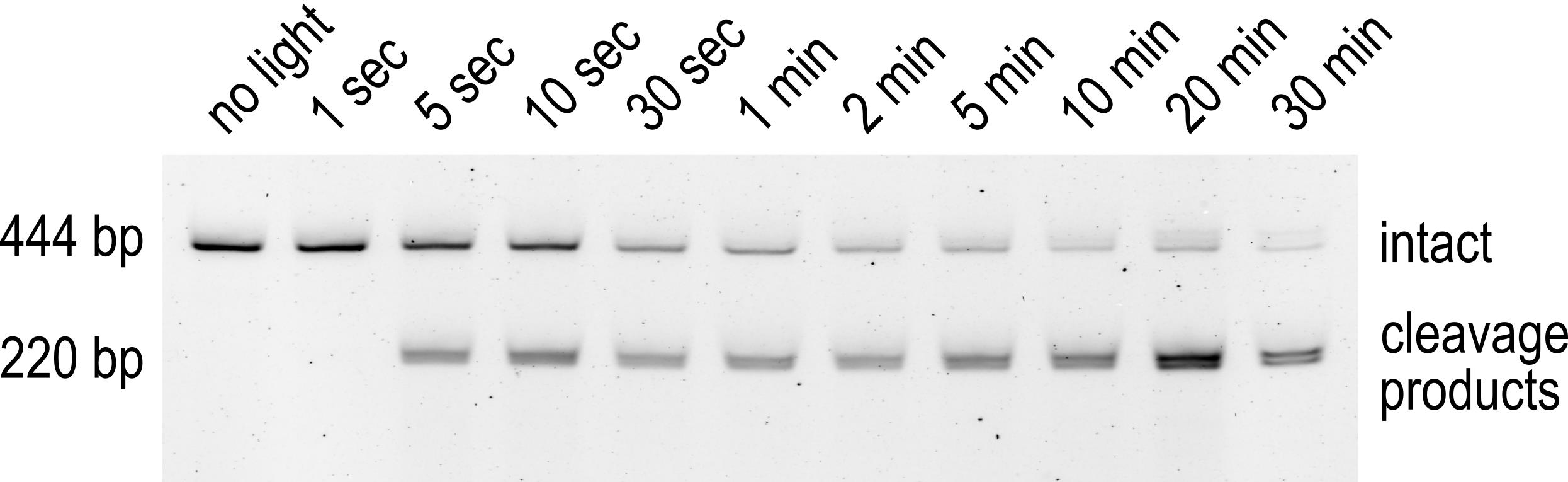
Figure 2. In vitro cleavage results. The top band migrating at 444 bp corresponds to the uncleaved PCR product of PPP1R2. The bottom band is formed by two unresolved bands, both migrating around 220 bp, which correspond to the cleavage products of the 444 bp top band. Each lane corresponds to either no light exposure or a different light exposure/incubation condition.
EMSA
Mix the following components together in order. Thoroughly mix NFW and NEBufferTM 3.1 before adding cgRNA and Cas9 and mixing again. Prepare 2 identical volumes, both in PCR tubes as samples {B} and {C}.
Component Volume NFW 8.1 μl NEBufferTM 3.1 1 μl 10 μM cgRNA targeting PPP1R2 0.5 μl 10 μM Cas9 0.4 μl Total 10 μl Prepare sample {A} by replacing Cas9 and cgRNA with water (negative control).
Leave on benchtop (room temperature) for 30 min to form RNP complex. Cover with aluminum film because photocaged gRNAs are light-sensitive.
Add 2 μl (~60 fmol) of PCR-amplified PPP1R2 target DNA to each of the three tubes. Mix well with pipette. For a highly efficient reaction, Cas9/cgRNA should be in at least 100-fold excess relative to target DNA. The target DNA should also be easily visible on an agarose gel with a gel imager.
Add 0.5 μl of Proteinase K to {C}, incubate at 55°C for 15 min.
Load 5 μl of each tube (in ascending order of time-duration) mixed with 15 μl water into each well of a 4% agarose E-Gel and run for 10 min. Add 100 bp ladder to the left-most lane. An alternative agarose gel electrophoresis system may also be used.
Visualize with TyphoonTM FLA 9000 using SYBR Gold setting. An alternative gel imager may also be used (Figure 3).
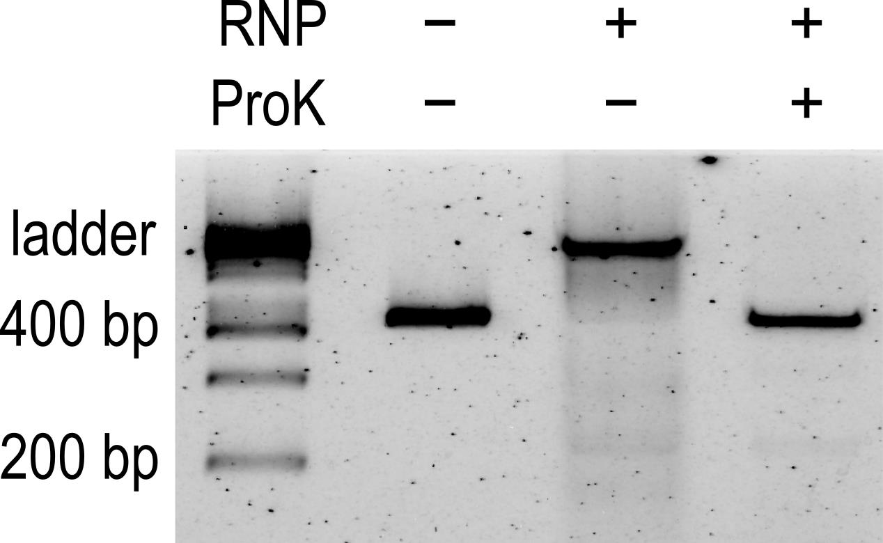
Figure 3. EMSA results. Lane 1 is the ladder; lane 2 is sample {A} (negative control without RNP); lane 3 is sample {B} (EMSA); lane 4 is sample {C} (Proteinase K digested sample). There is no cleavage product because the cgRNA was not exposed to any light, demonstrating that Cas9 with cgRNA binds to DNA even in the absence of light activation.
Acknowledgments
This work was supported by grants from the National Institutes of Health (R35 GM 122569 to T.H., and T32 GM 73009 and F30 CA 254160 to R. S. Z.) and the National Science Foundation (PHY 1430124 to T.H.). T.H. is an investigator of the Howard Hughes Medical Institute.
This protocol was derived from: Liu, Y., Zou, R. S., He, S., Nihongaki, Y., Li, X., Razavi, S., Wu, B. and Ha, T. (2020). Very fast CRISPR on demand. Science 368(6496): 1265-1269.
Competing interests
The authors and Johns Hopkins University have filed patent application PCT/US20/57256 on the method of spatiotemporal control of Cas9 activities via cgRNA.
References
- Adli, M. (2018). The CRISPR tool kit for genome editing and beyond. Nat Commun 9(1): 1911.
- Hellman, L. M. and Fried, M. G. (2007). Electrophoretic mobility shift assay (EMSA) for detecting protein-nucleic acid interactions. Nat Protoc 2(8): 1849-1861.
- Liu, Y., Zou, R. S., He, S., Nihongaki, Y., Li, X., Razavi, S., Wu, B. and Ha, T. (2020). Very fast CRISPR on demand. Science 368(6496): 1265-1269.
- Marraffini, L. A. (2015). CRISPR-Cas immunity in prokaryotes. Nature 526(7571): 55-61.
- Singh, D., Sternberg, S. H., Fei, J., Doudna, J. A. and Ha, T. (2016). Real-time observation of DNA recognition and rejection by the RNA-guided endonuclease Cas9. Nat Commun 7: 12778.
Article Information
Publication history
Accepted: May 13, 2021
Published: Sep 5, 2021
Copyright
© 2021 The Authors; exclusive licensee Bio-protocol LLC.
How to cite
Readers should cite both the Bio-protocol article and the original research article where this protocol was used:
- Zou, R. S., Liu, Y. and Ha, T. (2021). In vitro Cleavage and Electrophoretic Mobility Shift Assays for Very Fast CRISPR. Bio-protocol 11(17): e4138. DOI: 10.21769/BioProtoc.4138.
- Liu, Y., Zou, R. S., He, S., Nihongaki, Y., Li, X., Razavi, S., Wu, B. and Ha, T. (2020). Very fast CRISPR on demand. Science 368(6496): 1265-1269.
Category
Molecular Biology > DNA > DNA-protein interaction
Biological Sciences > Biological techniques
Do you have any questions about this protocol?
Post your question to gather feedback from the community. We will also invite the authors of this article to respond.
![]() Tips for asking effective questions
Tips for asking effective questions
+ Description
Write a detailed description. Include all information that will help others answer your question including experimental processes, conditions, and relevant images.