
- Submit a Protocol
- Receive Our Alerts
- EN

- Protocols

- Articles and Issues
- For Authors
- About
- Become a Reviewer

Isolation of Primary Cytotrophoblasts From Human Placenta at Term
(*contributed equally to this work) Published: Vol 11, Iss 19, Oct 5, 2021 DOI: 10.21769/BioProtoc.4185 Views: 1910
Reviewed by: Giusy TornilloDhiman Sankar PalAnonymous reviewer(s)
Original research article
The authors used this protocol in:
Nov 2020

Protocol Collections
Comprehensive collections of detailed, peer-reviewed protocols focusing on specific topics


Abstract
The placenta is a multifaceted organ, fulfilling critical functions for the fetus and the mother. Therefore, it is a critical regulator of the pregnancy, and its dysfunction leads to diseases, including fetal growth restriction and preeclampsia. Studying the placenta is a difficult task since its existence is transient, and its structure is specific to our species. In vitro differentiation of primary cytotrophoblast isolated from term human placenta has been widely used in the placental research field as it represents a reliable model to study cellular differentiation and function. Direct alternatives include trophoblastic cell lines, explants, and organoids, but this protocol, based on the separation of the cells on a Percoll gradient, presents the advantage of being relatively cheap and easy to perform in every research laboratory. Furthermore, the 2D culture is a flexible method that can be adapted to various experimental conditions (transfection, drug exposure, metabolic study, observations, etc.), allowing mechanistic explorations of cellular processes.
Keywords: TrophoblastBackground
Traditionally neglected by scientists, the placenta has long been regarded as a temporary organ with basic nutritional functions. Now, it is recognized as a complex and essential organ for mammalian reproduction, as aberrations in its formation and/or function have immediate consequences on pregnancy outcomes, as well as lifelong impacts on the health of the offspring (Malhotra et al., 2019). At term, the placenta has a discoid shape, with one side facing the amniotic cavity (the chorionic plate) and the other in contact with the endometrium (the decidual plate). Its microstructure is formed of villi that could be compared to multiple trees, which come from the branches of the fetal arteries (Turco and Moffett, 2019). Since these villi are directly in contact with the maternal blood, the human placenta is told hemochorial (Foidart et al., 1992). The interface between the maternal blood and the fetal vessels is called the syncytium. This unique layer constitutes the functional unit of the placenta and comes from the fusion of the mononuclear villous cytotrophoblasts (CTBs) to form multinucleated syncytiotrophoblasts (STBs). This process, known as syncytialization, takes place throughout the pregnancy and is critical for the formation and the functions of the placenta (Costa, 2016). On the other hand, the CTBs can also stay mononuclear but acquire an invasive capacity, especially during the first trimester. In this case, they are called extravillous trophoblasts (EVTs) and are crucial to the adaptations of the maternal arteries to pregnancy (Chang et al., 2018). In both cases, the study of CTBs differentiation represents one of the keys to improve our understanding of placental development and related diseases.
There are no perfect experimental models to investigate the human placenta as the differences between mammals are considerable, even in species with hemochorial placentas (e.g., rodents and non-human primates) (Roberts et al., 2016). Additionally, disorders such as preeclampsia, in which the primary defect is a failure in placentation, are found only in humans (Brosens et al., 2011). For these reasons, fundamental research on placental biology was initially based on ex vivo observations of histologic sections (Burton et al., 1999). In the last decades, various in vitro models based on human material were developed and characterized. However, each in vitro system has its benefits and limitations and should be carefully selected based on the trophoblast subtype, the gestational age of interest, and the type of experiment conducted (Colson et al., 2020b). In this context, primary human trophoblasts isolated from normal placenta at term and cultured in plastic cell culture dishes is an easy, cost-effective, and reproducible method to study specific molecular and genetic regulations in the placenta after the first trimester of gestation. This protocol is based on the method originally described by Kliman (Kliman et al., 1986) but was significantly improved by our team. The main advantages of this technique are its simplicity, its efficiency, and its cost. In addition, the method gives reproducible results as we have been able to characterize many independent cultures of several days (up to 12 days) over the last 10 years (Debieve et al., 2013; Depoix et al., 2013, 2016, 2018 and 2020; Colson et al., 2020a). Furthermore, the 2D culture is a flexible method that can be adapted to various experimental conditions (transfection, drug exposure, metabolic study, observations, etc.), allowing mechanistic explorations of cellular processes.
Limitations of this technique are the need for ethical permission, a good liaison with clinical staff, and the innate variability between samples. However, this latter limitation can be avoided by carefully selecting the placentas (similar gestational age, parity, and maternal condition) and repeated experiments. To overcome these difficulties, many research groups have turned to cell lines, including human embryonic stem cell-derived trophoblast cells (Amita et al., 2013), human trophoblast stem cells (Okae et al., 2018), and choriocarcinoma cell lines. Even if the first two cell lines require further characterization, their use is justified when the study is focused on early placentation as first-trimester trophoblasts are difficult to obtain. However, the use of choriocarcinoma cell lines such as JAR, JEG3, or BeWo to study CTB differentiation and functions poses real methodological difficulties. There are growing concerns about the purity, identity, and behavior of these common cell lines (Abbas et al., 2020). For instance, cAMP must be added to the culture medium to induce BeWo differentiation, while primary CTBs spontaneously fuse in vitro. Finally, genome-wide DNA methylation studies showed significant variability in comparison with primary cells, which is likely to contribute to differential expression profiles (Apps et al., 2011; Novakovic et al., 2011). Therefore, it becomes difficult to draw definitive conclusions when using such cell lines, which highlights the importance of validating findings by using primary trophoblasts.
Another alternative to primary human CTB cell culture is the use of placental explants from human placentas. Their culture is very easy and has the main advantage of keeping the CTBs in their complex tissue environment. It allows the comparison between placentas from normal pregnancies and pathological placentas (preeclampsia or fetal growth restriction) (Orendi et al., 2011). However, studies have shown that this technique impairs trophoblast viability and induces an unnatural oxidative stress (Di Santo et al., 2003; Goncalves et al., 2016). Since then, the use of explants in the study of placental biology must be restricted to comparisons with pregnancy-related diseases and must be limited in time. Finally, 3D cell culture models of human trophoblast cells that closely mimic the villous placenta were recently developed (Haider et al., 2018; Turco et al., 2018). These organoids, composed of a layer of CTBs surrounding a core of STBs, can also be differentiated into EVTs. These models undoubtedly represent a breakthrough in the understanding of placental biology but require specific technical skills and a consequent budget. For these reasons, we are convinced that primary human term CTB cultures present undeniable advantages that still justify their place in the placental field.
Materials and Reagents
Disposable
Percoll (1.130 ± 0.005 g/ml) (GE Healthcare Bio-Sciences AB, catalog number: GE17-0891-01). Keep it sterile and conserve at 4°C
10× Hank’s Buffered Saline Solution w/o Ca, w/o Mg, w/o Phenol red (Gibco, Thermo Fisher Scientific, catalog number: 14175053). Keep it sterile and conserve at 4°C
1× Hank’s Buffered Saline Solution w/o Ca, w/o Mg, w/o Phenol red (Gibco, Thermo Fisher Scientific, catalog number: 14175095). Keep it sterile and conserve at 4°C
Dulbecco's modified Eagle medium (Thermo Fisher Scientific, catalog number: 12440061). Keep it sterile and conserve at 4°C
Iscove’s Modified Dulbecco Medium (Gibco, Thermo Fisher Scientific, catalog number: 12440061). Keep it sterile and conserve at 4°C
Gentamicin 50 mg/ml (Gibco, Thermo Fisher Scientific, catalog number: 15750037). Keep it sterile and conserve at RT
Sterile heat-inactivated Fetal Bovine Serum (Gibco, Thermo Fisher Scientific, catalog number: 10082147). Keep it sterile. Aliquote and store at -20°C
Dulbecco's phosphate-buffered saline (DPBS) (Gibco, Thermo Fisher Scientific, catalog number: 14190359). Keep it sterile and conserve at 4°C
Lyophilized dispase II (Roche Diagnostics, catalog number: 4942078001). Store at 4°C
DNAse I, grade II (Roche Diagnostics, catalog number: 10104159001). Resuspend at 10 mg/ml and store at -20°C
4 L to 8 L NaCl 0.9% (Carl Roth GmbH + Co. KG, catalog number: 9265.2). Keep it sterile and conserve at 4°C
Nunc 200 ml conical centrifuge tubes (Thermo Fisher Scientific, catalog number: 376813)
15 ml centrifuge tubes (Greiner Bio-One International GmbH, catalog number: 188261)
50 ml centrifuge tubes (Greiner Bio-One International GmbH, catalog number: 227261)
5 ml serological pipets (Greiner Bio-One International GmbH, catalog number: 606180)
10 ml serological pipets (Greiner Bio-One International GmbH, catalog number: 607180)
20 ml syringes (Greiner Bio-One International GmbH, catalog number: 474203)
50 ml syringes (Greiner Bio-One International GmbH, catalog number: 475203)
Blunt-end 16 G needles (VWR International, catalog number: POPR7938)
Corning 40 µm disposable cell strainer (Corning, catalog number: 431750)
Plastics for cell culture. Coating with collagen or laminin is not necessary, but better results can be obtained by using pretreated plastics such as the Advanced TC products (Greiner Bio-One International GmbH, catalog number: 665980)
Non-Disposable Sterilized Materials
One dish stainless-steel (Carl Roth GmbH + Co. KG, catalog number: 0376.1)
One pair of big scissors (Carl Roth GmbH + Co. KG, catalog number: YE80.1)
One pair of little scissors (Carl Roth GmbH + Co. KG, catalog number: HEY6.1)
One tweezer straight (Carl Roth GmbH + Co. KG, catalog number: 2802.1)
One big round sieve (Carl Roth GmbH + Co. KG, catalog number: 8101.1)
One little round sieve (Carl Roth GmbH + Co. KG, catalog number: 8096.1)
One funnel (Carl Roth GmbH + Co. KG, catalog number: 0166.1)
One flat glass recipient (Carl Roth GmbH + Co. KG, catalog number: HX16.1)
One 500 m baffled flasks with screw closure (Carl Roth GmbH + Co. KG, catalog number: PK17.1)
One 500 ml baffled flasks, straight neck (Carl Roth GmbH + Co. KG, catalog number: LY96.1)
One 1 L beaker (Carl Roth GmbH + Co. KG, catalog number: X695.1)
One 2 L beaker (Carl Roth GmbH + Co. KG, catalog number: X696.2)
One stainless-steel test sieve, pore size 200 µm (Retsch, catalog number: 60.106.000200)
One stainless-steel test sieve, pore size 100 µm (Retsch, catalog number: 60.106.000100)
One stainless-steel test sieve, pore size 40 µm (Retsch, catalog number: 60.106.000040)
Equipment
Pipet controller
Vacuum machine
Bucket
Refrigerated centrifuge at 4°C with adaptors for 15 ml tubes, 50 ml tubes, and 200 ml centrifugation tubes
Stand with a clamp
Surgical lamp (cold light)
Laminar flow hood
Atmosphere-controlled humidified incubator
Scale
Shaking water bath at 37°C
Fluorescence microscope and camera
Procedure
See the video 1 joined to this protocol to get a complete overview of the procedure.

Discontinuous Percoll Gradient Preparation
Prepare all the solutions and the gradient under a laminar flow hood.
Prepare one solution of 90% Percoll by mixing 19.8 ml of stock Percoll (100%) with 2.2 ml of 10× HBSS.
Prepare the following dilutions in 15 ml tubes as indicated in Table 1. Briefly centrifuge the solutions and store them at 4°C. The Percoll dilutions can be prepared several days before the isolation procedure.
Table 1. Preparation of the Percoll dilutions for the discontinuous gradient
% 90% Percoll (ml) 1× HBSS (ml) 70 2.8 0.8 65 2.6 1 60 2.4 1.2 55 2.2 1.4 50 2 1.6 45 1.8 1.8 40 1.6 2 35 1.4 2.2 30 1.2 2.4 25 1 2.6 20 0.8 2.8 15 0.6 3 10 0.4 3.2 5 0.2 3.4 In a 50 ml tube, delicately drop off 3 ml of each dilution, layer-by-layer, starting with the 70% dilution. To facilitate the process, use a 5 ml serological pipet with the pipet controller set on the passive mode and let the solution descend very slowly.
Keep the gradient at 4°C for optimal separation of the cells. It must be freshly prepared the day of the isolation.
Placenta Collection
Only include placentas retrieved from strictly normal singleton pregnancies at term (37-41 weeks of gestation), without any fetal (growth restriction, malformations, genomic abnormalities, or infection), placental (unique umbilical artery, placenta praevia, or placenta accreta), or maternal (chronic diseases, gestational diabetes, preeclampsia, medications, alcohol, or drugs) complications.
Collect material on ice immediately after cesarean section and proceed with digestion within one hour.
Tissue Preparation
Put the placenta in the stainless steel container and roughly remove the amniotic membranes with the pair of big scissors.
Cut the placenta into 2 × 2 cm pieces and put them in the 2 L beaker (Figure 1A).
Wash the tissue by adding 500 ml of NaCl 0.9%. Mix well and drain the pieces in the big sieve placed on the bucket (Figure 1B).
Repeat this operation until the wash solution remains clear (4 L to 8 L needed).
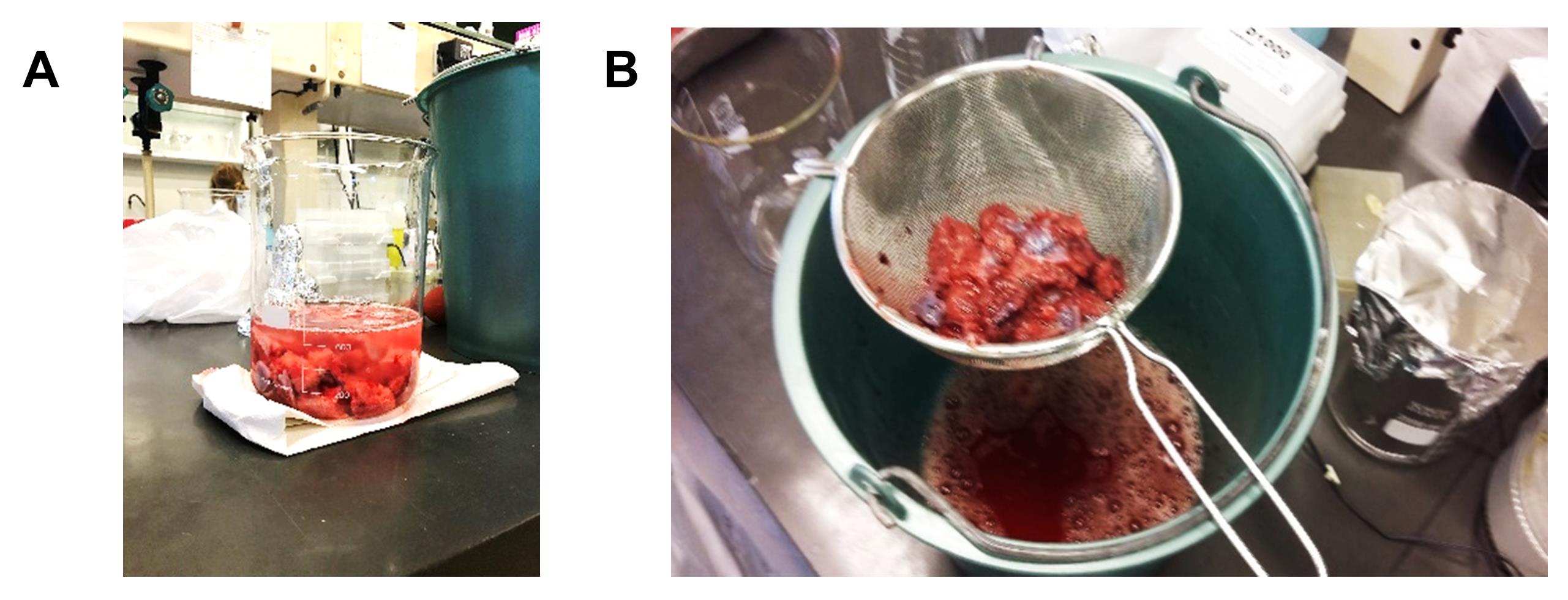
Figure 1. Washing step. A. After cutting the placenta into pieces, put them in the 2 L beaker and add 500 ml of NaCl 0.9%. B. Shake the beaker vigorously and drain the tissue in the sieve. Repeat this operation several times. After 4 to 8 L of NaCl 0.9%, the pieces must turn light red, and the wash solution must remain clear.Place the pieces in the glass container and delicately dry the tissue with paper towels (Figure 2A).
With the pair of little scissors and the tweezer, remove the big blood vessels (Figure 2B), the chorionic plate and the remaining amniotic membranes (the white side) (Figure 2C and 2D), the decidua (the pearly opposite side), and the blood clots to facilitate the digestion and to reduce the amount of debris.
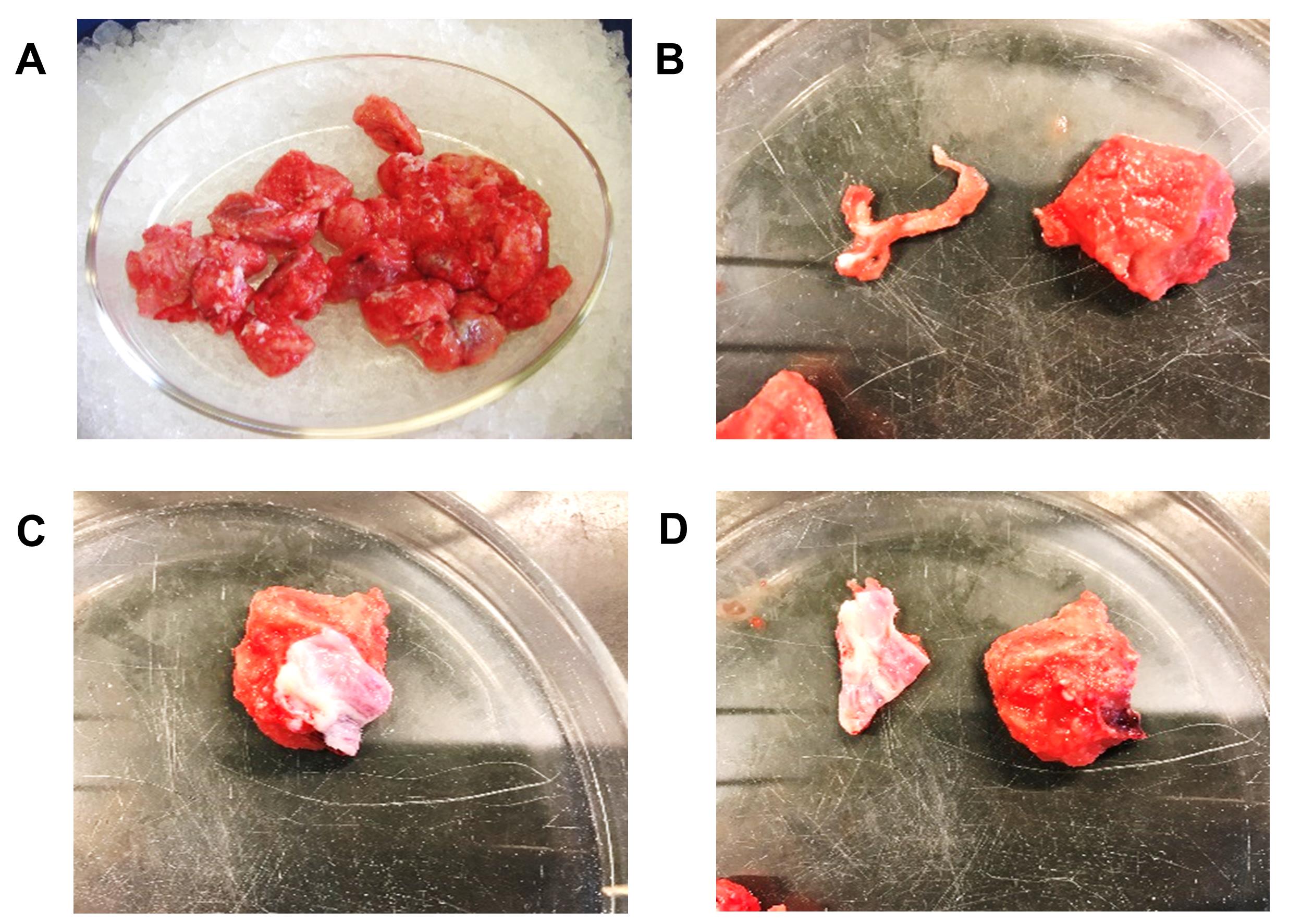
Figure 2. Cleaning step. Prepare the pieces for digestion. A. After drying with paper towels, B. Remove big blood vessels and C-D. the chorionic plate. This step can be time-consuming, but reducing the amount of debris improves digestion and protocol yield.Using the scale, weigh 72 g of tissue and chop the fragments with the pair of big scissors (Figure 3A).
Enzymatic Digestion
In the water bath, preheat 300 ml of 1× HBSS in the 500 ml baffled flasks with screw closure.
Dissolve 720 units of lyophilized dispase I in the preheated 1× HBSS to obtain a solution at 2.4 units/ml.
Add the finely minced placental tissue to the dispase solution and incubate 1 h in the water bath under strong shaking (Figure 3B).
Add 1.2 ml of DNAse I 10 mg/ml to the digested tissue to obtain a final concentration of 100 units/ml.
Incubate in the water bath under strong shaking for another 15 min.
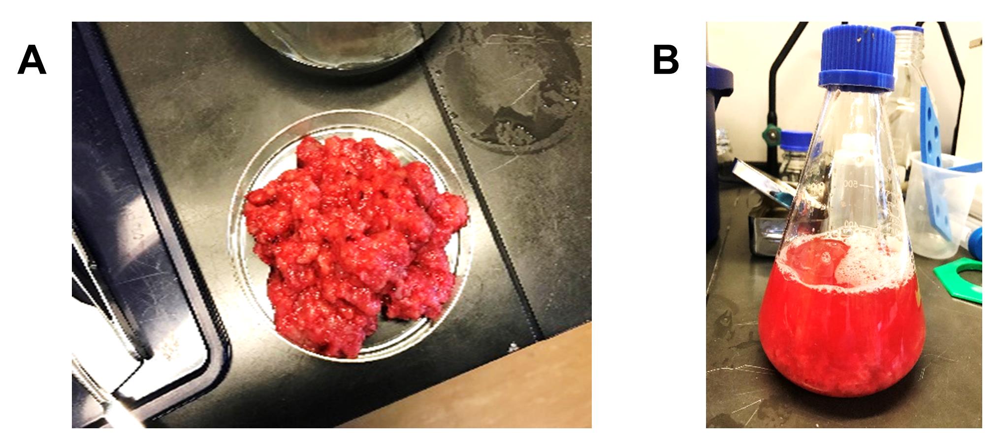
Figure 3. Digestion step. Dissolve 720 units of dispase I in 300 ml of preheated HBSS. A. Chop the pieces with the pair of big scissors and transfer the pulp to the baffled flask. B. Incubate the mixture 1 h in the water bath under strong shaking. Add predissolved DNAse I and incubate for another 15 min.
Cell Isolation and Purification
Prefilter the suspension using in the little sieve placed on the 1 L beaker (Figure 4A).
Fix the three test sieves in decreasing order (pore diameter: 200 µm-100 µm-40 µm) on the stand with the clamp.
Place the stainless steel funnel and the 500 ml baffled flasks with a straight neck below the filters.
Let the suspension pass through the filtration system. If the filtration is difficult, agitate the test sieves to move debris and draw air. Alternatively, a porcelain spoon can also be used to remove blocking dirt (Figure 4B).
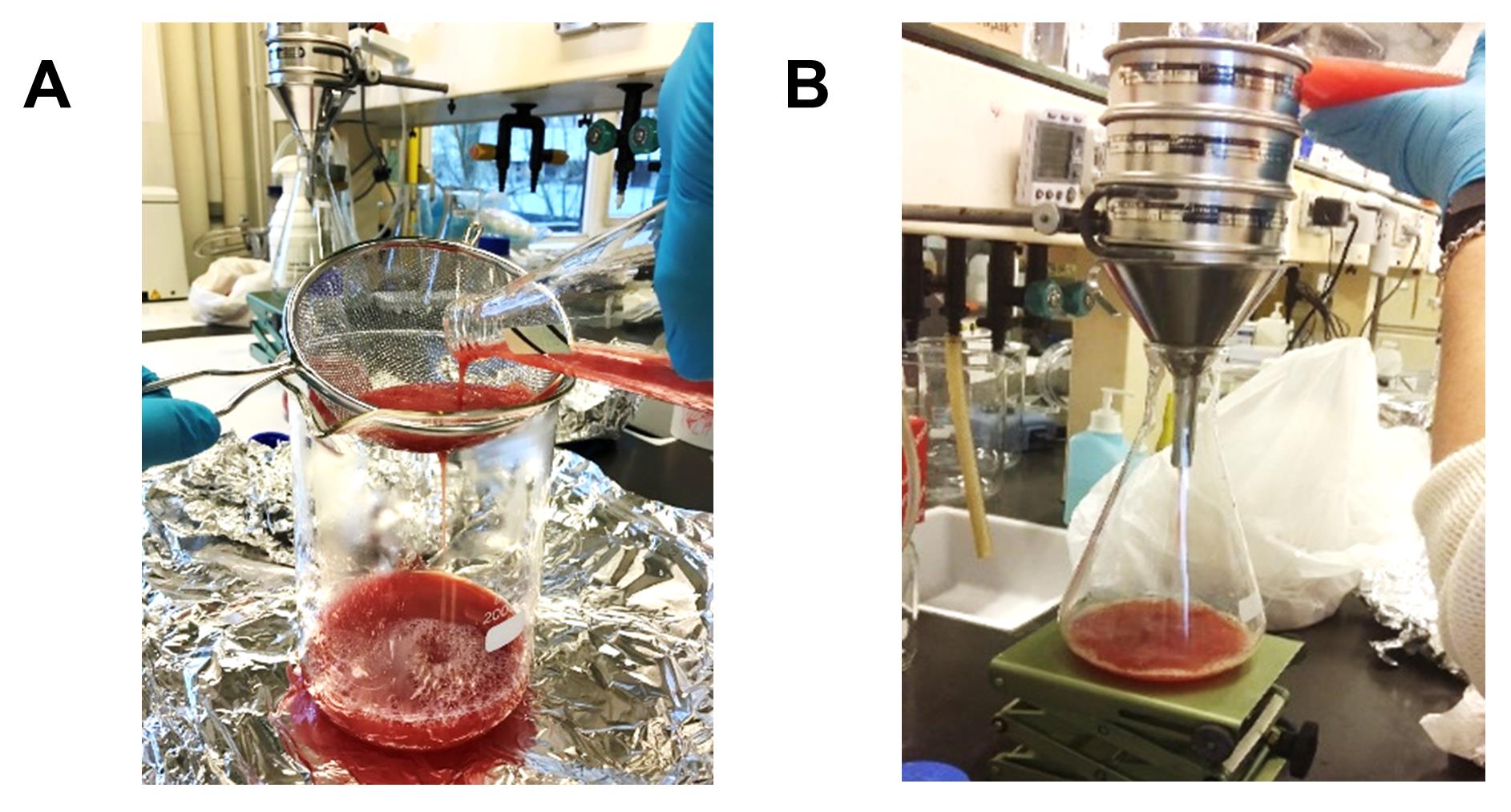
Figure 4. Filtration step. A. After the digestion, the suspension is first prefiltered by using the little sieve. B. The suspension is then passed through the three test sieves, fixed on the stand by decreasing order. The filtrate is finally centrifuged; the pellet contains red blood cells, lymphocytes, debris, and cytotrophoblasts.Divide the filtrate into two centrifugation bottles and complete with IMDM or DMEM medium supplemented with gentamicin (final concentration 50 µg/ml). This will be referred to as “wash medium” in the rest of the procedure.
Centrifuge 20 min at 500 × g.
Aspirate the supernatant. The pellet contains red blood cells, lymphocytes, debris, and cytotrophoblasts. Be careful not to aspirate the pellet, as it is friable at this step of the procedure.
Resuspend the pellet with 10 ml of wash medium and transfer to a new 50 ml tube.
Complete to 50 ml with wash medium and centrifuge 5 min at 500 × g.
Aspirate the supernatant and resuspend the pellet in 6 ml of wash medium.
Delicately lay down these 6 ml on the surface of the gradient with the pipet controller (Figure 5A and 5B).
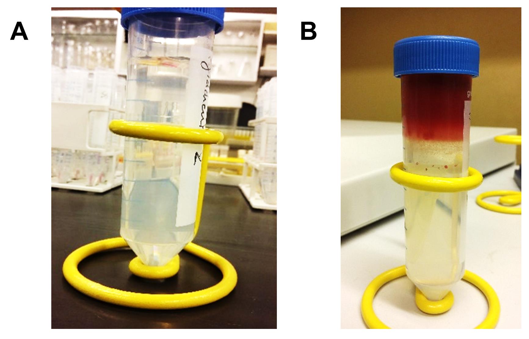
Figure 5. Gradient step. The 6 ml contains a mix of debris, red blood cells, lymphocytes, and cytotrophoblasts. The discontinuous Percoll gradient allows separating the cell type by density. A. The gradient was freshly prepared on the day of isolation and stored at 4°C until use. B. After the filtration step, the cell suspension is meticulously deposited at the surface of the gradient. It is then centrifuged to separate the cells between the different layers.Centrifuge 20 min at 1,200 × g without brake.
After centrifugation, place the gradient in front of the surgical lamp (cold light) to visualize the cells. The cytotrophoblasts form a thick layer localized between 12.5 ml and 25 ml, which corresponds to a density of 1.048-1.062 g/ml (layers 45%-40%-35%) (Figure 6). This has been previously determined with density marker beads. The debris must be localized in the low-density layers, whereas the lymphocytes can be visualized below the cytotrophoblasts. Some red blood cells may be observed within the cytotrophoblast layer, but the majority of them should be located at the bottom of the tube.
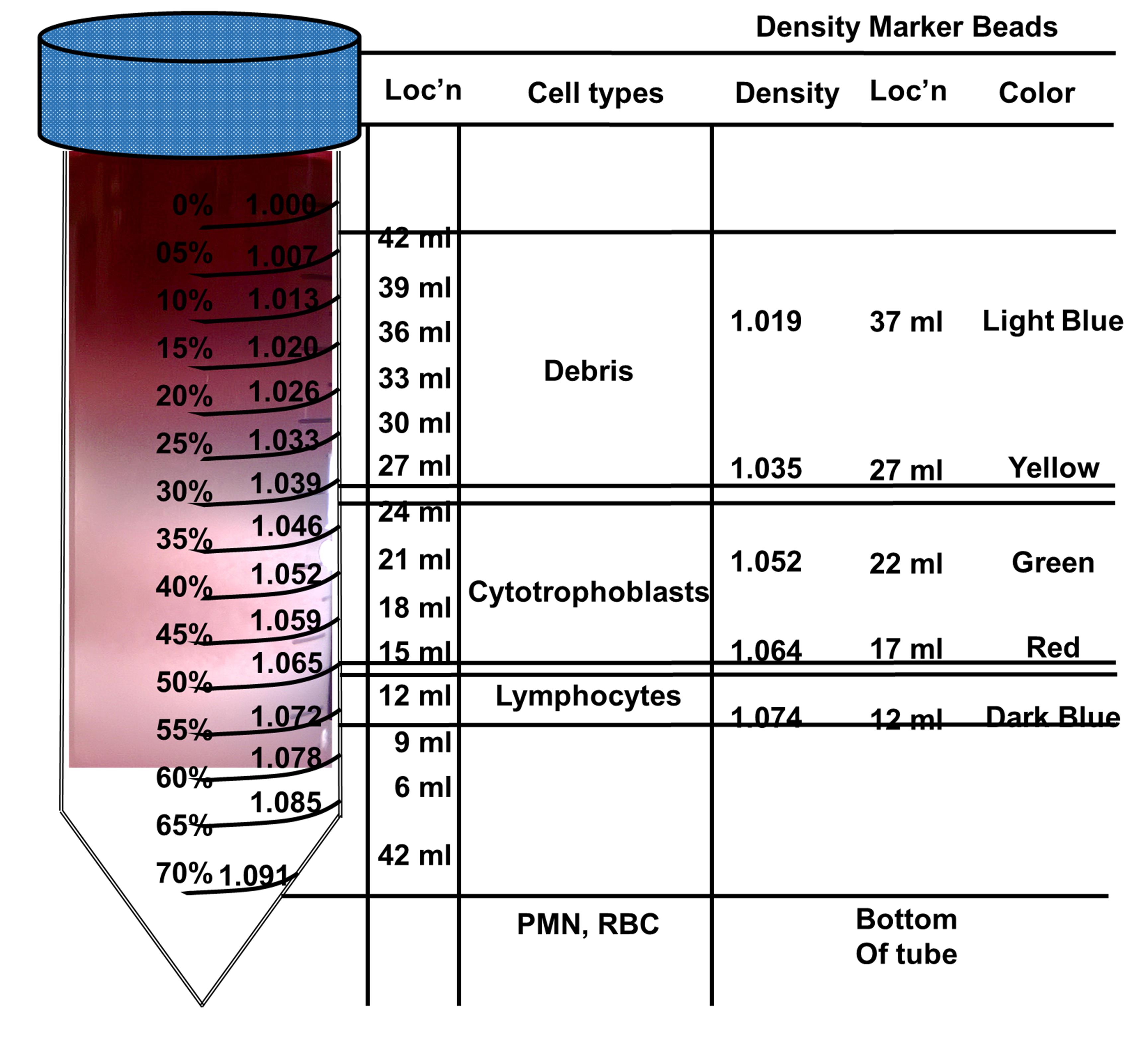
Figure 6. The discontinuous Percoll gradient after centrifugation. The cells form a compact cloud between 12.5 and 20 ml. The debris (red) are above, while the red blood cells form a pellet at the bottom of the tube. Some lymphocytes can be observed around the 12 ml graduation.Remove by aspiration the upper layers corresponding to the debris with a 50 ml syringe and a blunt-end 15 G needle.
With a new 20 ml syringe and the other blunt-end 15 G needle, aspirate the cell layer corresponding to the cytotrophoblasts and put them in a new 50 ml tube.
Complete the volume to 50 ml with wash medium and centrifuge 5 min at 300 × g.
The cytotrophoblasts form a white pellet at the bottom of the tube. Some red blood cells can be occasionally observed.
Remove the supernatant and thoroughly resuspend the pellet with 6 ml of 0.05% trypsin.
Manually agitate the tube 1 min in the water bath and stop the reaction by immediately placing the tube on ice.
Add wash medium rapidly to 50 ml and centrifuge 5 min at 300 × g.
Remove the supernatant and resuspend the cell pellet in 10 ml of wash medium.
Filtrate the solution with a 40-µm disposable cell strainer.
Complete the volume to 50 ml with wash medium and proceed to counting.
Cell Counting and Culture
Dilute 60 µl of the cell suspension in 260 µl of wash medium.
Add 100 µl of 0.4% Trypan blue.
Count the cells with a hemocytometer (Bürker) and determine the percentage of viability.
Centrifuge the cell suspension 5 min at 300 × g.
Resuspend the pellet in complete culture medium (IMDM-10% FBS, gentamicin 50 μg/ml). From now, the cytotrophoblasts must be cultured with IMDM.
Plate the cells at a density of 4.0 × 10⁵ cells/cm2 to achieve immediate confluence (for example, 1.5 × 10⁶ cells/well in a 12-well plate).
Place the cells in an atmosphere-controlled humidified incubator under 21% O2-5% O2.
The day after, wash the cells with preheated 1× DPBS to remove debris, syncytial knots, and unattached cells.
Aspirate the DPBS and add preheated culture medium. Change the medium every 24 h.
The cells can be cultured for several days (up to 21 days). Some syncytiotrophoblasts can already be observed after 24 h, and the maximum syncytialization is achieved 72-96 h after isolation.
We advise checking the purity of the culture the first few times the protocol is performed (Supplementary file).
Acknowledgments
This protocol was initially developed by Frédéric Debiève and optimized by Christophe Depoix and Arthur Colson during the last few years with the technical support of Séverine Gonze. The authors are particularly grateful to medical doctors and midwives of the department of obstetrics of the Saint-Luc University Hospital for providing access to fresh placentas. The development of this technique was fully supported by the charity Fetus for Life. AC is a PhD Fellow of the Belgian Fonds pour la Formation à la Recherche dans l'Industrie et dans l'Agriculture (FRIA).
Competing interests
The authors declare no competing interest.
Ethics
The work performed in our lab was conducted with the approval of the Ethical Committee of the Catholic University of Louvain and the Saint-Luc University Hospital (Brussels, Belgium) under the reference 2018/23OCT/397. Informed consent was obtained from all patients.
References
- Abbas, Y., Turco, M. Y., Burton, G. J. and Moffett, A. (2020). Investigation of human trophoblast invasion in vitro. Hum Reprod Update 26(4): 501-513.
- Amita, M., Adachi, K., Alexenko, A. P., Sinha, S., Schust, D. J., Schulz, L. C., Roberts, R. M. and Ezashi, T. (2013). Complete and unidirectional conversion of human embryonic stem cells to trophoblast by BMP4. Proc Natl Acad Sci U S A 110(13): E1212-1221.
- Apps, R., Sharkey, A., Gardner, L., Male, V., Trotter, M., Miller, N., North, R., Founds, S. and Moffett, A. (2011). Genome-wide expression profile of first trimester villous and extravillous human trophoblast cells. Placenta 32(1): 33-43.
- Brosens, I., Pijnenborg, R., Vercruysse, L. and Romero, R. (2011). The "Great Obstetrical Syndromes" are associated with disorders of deep placentation. Am J Obstet Gynecol 204(3): 193-201.
- Burton, G. J., Jauniaux, E. and Watson, A. L. (1999). Maternal arterial connections to the placental intervillous space during the first trimester of human pregnancy: the Boyd collection revisited. Am J Obstet Gynecol 181(3): 718-724.
- Chang, C. W., Wakeland, A. K. and Parast, M. M. (2018). Trophoblast lineage specification, differentiation and their regulation by oxygen tension. J Endocrinol 236(1): R43-R56.
- Colson, A., Depoix, C. L., Baldin, P., Hubinont, C., Sonveaux, P. and Debieve, F. (2020a). Hypoxia-inducible factor 2 alpha impairs human cytotrophoblast syncytialization: New insights into placental dysfunction and fetal growth restriction. FASEB J 34(11): 15222-15235.
- Colson, A., Sonveaux, P., Debieve, F. and Sferruzzi-Perri, A. N. (2020b). Adaptations of the human placenta to hypoxia: opportunities for interventions in fetal growth restriction. Hum Reprod Update 27(3): 531-569.
- Costa, M. A. (2016). Scrutinising the regulators of syncytialization and their expression in pregnancy-related conditions. Mol Cell Endocrinol 420: 180-193.
- Debieve, F., Depoix, C., Gruson, D. and Hubinont, C. (2013). Reversible effects of oxygen partial pressure on genes associated with placental angiogenesis and differentiation in primary-term cytotrophoblast cell culture. Mol Reprod Dev 80(9): 774-784.
- Depoix, C., Barret, L. A., Hubinont, C. and Debieve, F. (2013). Viability of primary term cytotrophoblast cell culture in normoxia and hypoxia. Mol Hum Reprod 19(1): 29-34.
- Depoix, C. L., Colson, A., Hubinont, C. and Debieve, F. (2020). Impaired vascular endothelial growth factor expression and secretion during in vitro differentiation of human primary term cytotrophoblasts. Angiogenesis 23(2): 221-230.
- Depoix, C. L., Flabat, O., Debieve, F. and Hubinont, C. (2016). HIF1A and EPAS1 mRNA and protein expression during in vitro culture of human primary term cytotrophoblasts and effect of oxygen tension on their expression. Reprod Biol 16(3): 203-211.
- Depoix, C. L., Haegeman, F., Debieve, F. and Hubinont, C. (2018). Is 8% O2 more normoxic than 21% O2 for long-term in vitro cultures of human primary term cytotrophoblasts? Mol Hum Reprod 24(4): 211-220.
- Di Santo, S., Malek, A., Sager, R., Andres, A. C. and Schneider, H. (2003). Trophoblast viability in perfused term placental tissue and explant cultures limited to 7-24 hours. Placenta 24(8-9): 882-894.
- Foidart, J. M., Hustin, J., Dubois, M. and Schaaps, J. P. (1992). The human placenta becomes haemochorial at the 13th week of pregnancy. Int J Dev Biol 36(3): 451-453.
- Goncalves, J. M., Casart, Y. C. and Camejo, M. I. (2016). Nitric oxide and oxidative stress in placental explant cultures. Syst Biol Reprod Med 62(1): 11-16.
- Haider, S., Meinhardt, G., Saleh, L., Kunihs, V., Gamperl, M., Kaindl, U., Ellinger, A., Burkard, T. R., Fiala, C., Pollheimer, J., Mendjan, S., Latos, P. A. and Knofler, M. (2018). Self-Renewing Trophoblast Organoids Recapitulate the Developmental Program of the Early Human Placenta. Stem Cell Reports 11(2): 537-551.
- Kliman, H. J., Nestler, J. E., Sermasi, E., Sanger, J. M. and Strauss, J. F., 3rd (1986). Purification, characterization, and in vitro differentiation of cytotrophoblasts from human term placentae. Endocrinology 118(4): 1567-1582.
- Malhotra, A., Allison, B. J., Castillo-Melendez, M., Jenkin, G., Polglase, G. R. and Miller, S. L. (2019). Neonatal Morbidities of Fetal Growth Restriction: Pathophysiology and Impact. Front Endocrinol (Lausanne) 10: 55.
- Novakovic, B., Gordon, L., Wong, N. C., Moffett, A., Manuelpillai, U., Craig, J. M., Sharkey, A. and Saffery, R. (2011). Wide-ranging DNA methylation differences of primary trophoblast cell populations and derived cell lines: implications and opportunities for understanding trophoblast function. Mol Hum Reprod 17(6): 344-353.
- Okae, H., Toh, H., Sato, T., Hiura, H., Takahashi, S., Shirane, K., Kabayama, Y., Suyama, M., Sasaki, H. and Arima, T. (2018). Derivation of Human Trophoblast Stem Cells. Cell Stem Cell 22(1): 50-63 e56.
- Orendi, K., Kivity, V., Sammar, M., Grimpel, Y., Gonen, R., Meiri, H., Lubzens, E. and Huppertz, B. (2011). Placental and trophoblastic in vitro models to study preventive and therapeutic agents for preeclampsia. Placenta 32 Suppl: S49-54.
- Roberts, R. M., Green, J. A. and Schulz, L. C. (2016). The evolution of the placenta. Reproduction 152(5): R179-189.
- Turco, M. Y., Gardner, L., Kay, R. G., Hamilton, R. S., Prater, M., Hollinshead, M. S., McWhinnie, A., Esposito, L., Fernando, R., Skelton, H., et al. (2018). Trophoblast organoids as a model for maternal-fetal interactions during human placentation. Nature 564(7735): 263-267.
- Turco, M. Y. and Moffett, A. (2019). Development of the human placenta. Development 146(22): dev163428.
Article Information
Publication history
Accepted: Jul 12, 2021
Published: Oct 5, 2021
Copyright
© 2021 The Authors; exclusive licensee Bio-protocol LLC.
How to cite
Colson, A., Depoix, C. L., Hubinont, C. and Debiève, F. (2021). Isolation of Primary Cytotrophoblasts From Human Placenta at Term. Bio-protocol 11(19): e4185. DOI: 10.21769/BioProtoc.4185.
Category
Developmental Biology > Reproduction
Cell Biology > Cell isolation and culture > Cell isolation
Do you have any questions about this protocol?
Post your question to gather feedback from the community. We will also invite the authors of this article to respond.
![]() Tips for asking effective questions
Tips for asking effective questions
+ Description
Write a detailed description. Include all information that will help others answer your question including experimental processes, conditions, and relevant images.