
- Submit a Protocol
- Receive Our Alerts
- EN

- Protocols

- Articles and Issues
- For Authors
- About
- Become a Reviewer

Establishment of an in vitro Differentiation and Dedifferentiation System of Rat Schwann Cells
Published: Vol 13, Iss 5, Mar 5, 2023 DOI: 10.21769/BioProtoc.4631 Views: 415
Reviewed by: Gal HaimovichPhilipp WörsdörferAnonymous reviewer(s)
Original research article
The authors used this protocol in:

Jan 2022

Protocol Collections
Comprehensive collections of detailed, peer-reviewed protocols focusing on specific topics


Related protocols
Abstract
In the peripheral nervous system, Schwann cells are the primary type of glia; their in vitro differentiation and dedifferentiation system has not been described in detail in the literature. Thus, an in vitro differentiation and dedifferentiation system of rat Schwann cells is described in this protocol. These cultures and systems may be used to investigate the morphological and biochemical effects of drug interventions or lentivirus-mediated gene transfer on Schwann cells during differentiation or dedifferentiation.
Graphical abstract

Background
Schwann cells are the primary glial cells of the peripheral nervous system. During axonal sorting and myelination in the peripheral nerves, Schwann cells originate from neural crest cells that differentiate into mature phenotypes (Cristobal and Lee, 2022). Moreover, Schwann cells’ incredible plasticity is one of the most important characteristics following nerve damage or demyelination (Nocera and Jacob, 2020). Schwann cells undergo dedifferentiation after injury and redifferentiate to promote nerve regeneration and complete functional recovery (Jessen and Mirsky, 2019). Therefore, it is important to study Schwann cells’ differentiation and dedifferentiation status to understand their role in nerve development and injury. As reported previously, dibutyryl adenosine 3',5'-cyclic monophosphate (dbcAMP) induces Schwann cells to acquire a differentiated phenotype (Yamauchi et al., 2011). After stimulation with 1 mM dbcAMP, Schwann cells from rats change from bipolar or tripolar to flat within 24 h. By contrast, mouse Schwann cells retain their bipolar or tripolar morphology after dbcAMP treatment (Arthur-Farraj et al., 2011). However, the details of how Schwann cells acquire a differentiated or dedifferentiated phenotype in vitro remain poorly understood. Herein, we established a detailed system of differentiation and dedifferentiation of Schwann cells. In order to clearly distinguish morphological changes, we performed the differentiation and dedifferentiation assay using rat Schwann cells. In brief, the first step was to obtain Schwann cells from the spinal nerves of rats based on a previous study (Wen et al., 2017). During the second step, differentiation and dedifferentiation assays were conducted. Schwann cells were purified and passaged, and experiments were conducted on the third passage. Finally, Schwann cell status was determined by morphological examination, western blotting analysis, and immunofluorescence detection.
Materials and Reagents
Cell culture dish (3.5 and 6 cm) (JET BIOFILT, catalog numbers: CD000035, TCD-010-060)
50 mL centrifuge tubes (Corning, catalog number: 430828)
15 mL centrifuge tubes (Corning, catalog number: 430790)
1.5 mL tube (JET BIOFILT, catalog number: CFT000015)
Polyvinylidene fluoride membrane (PVDF) (Bio-Rad, catalog number: 1620177)
Cell glass coverslips (diameter: 12 mm, thickness: 0.13–0.17 mm) (Fisherbrand, catalog number: FIS12-545-80)
Neonatal Sprague-Dawley (SD) rat [postnatal 1–2 days (P1–P2)]
Distilled water
75% ethanol
Phosphate buffered saline (PBS) (Gibco, catalog number: 10010023)
Poly-L-lysine hydrobromide (PLL) (Sigma-Aldrich, catalog number: P1274)
0.25% trypsin-EDTA (Gibco, catalog number: 25200072)
Fetal bovine serum (FBS) (Corning, catalog number: 35-076-CV)
DMEM/F12 (Gibco, catalog number: 11330057)
Cytosine arabinoside (Ara-C) (Sigma-Aldrich, catalog number: C1768)
Recombinant human heregulin β-1 (PeproTech, catalog number: 100-03)
Forskolin (Sigma-Aldrich, catalog number: F6886)
4% paraformaldehyde (PFA) (Biosharp, catalog number: BL539A)
Dimethyl sulfoxide (DMSO), suitable for cell culture (Beyotime, catalog number: ST038)
Dibutyryl adenosine 3',5'-cyclic monophosphate (dbcAMP) (Sigma-Aldrich, catalog number: D0627)
Triton X-100 (Sigma-Aldrich, catalog number: V900502)
Tween-20 (Sigma-Aldrich, catalog number: P1379)
Phalloidin (Abcam, catalog number: ab176759)
RIPA lysis buffer (FUDE Biological Technology, catalog number: FD009)
Rabbit monoclonal (EP1039Y) anti-p75 (Abcam, catalog number: ab52987)
Rabbit anti-Krox20 (Novus Biologicals, catalog number: 13491-1-AP)
Mouse monoclonal anti-c-Jun (BD Biosciences, catalog number: 610326)
Mouse monoclonal (9-9-3) anti-Sox2 (Abcam, catalog number: ab79351)
Alexa Fluor® 488 goat anti-mouse IgG (H+L) (Thermo Fisher Scientific, catalog number: A11001)
Alexa Fluor® 568 goat anti-rabbit IgG (H+L) (Thermo Fisher Scientific, catalog number: A11011)
4’,6-diamidino-2’-phenylindole (DAPI) (Sigma-Aldrich, catalog number: D8417)
Penicillin–streptomycin (Gibco, catalog number: 15140-122)
Omni-ECLTM Light Chemiluminescence kit (EpiZyme, catalog number: SQ201)
10% dodecyl sulfate sodium salt-polyacrylamide gel (EpiZyme, catalog number: PG112)
5% non-fat milk (Solarbio, catalog number: D8340)
Tris-buffered saline (Sigma-Aldrich, catalog number: 93318)
Horseradish peroxidase (HRP)-conjugated secondary antibody (Invitrogen, catalog numbers: 31430, 31460)
1,000× Ara-C (10 mM in distilled water, see Recipes)
1,000× dbcAMP (1,000 mM in DMSO, see Recipes)
30 mM forskolin stock solution (see Recipes)
Complete growth medium of rat Schwann cells (see Recipes)
10% FBS (see Recipes)
3% FBS (see Recipes)
1% FBS (see Recipes)
1 mM dbcAMP (see Recipes)
0.1% Triton X-100 (see Recipes)
5% gelatin (see Recipes)
PBST (see Recipes)
Blocking buffer (see Recipes)
Equipment
Pipettes (Thermo Fisher Scientific, 10, 200, and 1,000 μL)
CO2 incubator (Thermo Fisher Scientific, model: Heracell 150i)
Surgical scissors and forceps (Shenzhen RWD, catalog numbers: S14014 and F12029-09)
Spring scissors (Shenzhen RWD, catalog number: S11001)
Superfine forceps (Shenzhen RWD, catalog number: F13002)
Stereomicroscope (Jiangnan Novel, model: SZ6060)
Centrifuge (Eppendorf, model: Micro21)
Phase contrast microscope (Zeiss, model: Primovert)
Fluorescence microscope (Zeiss, model: Axio Imager A2)
Software
ImageJ (Version 1.8.0, https://imagej.en.softonic.com/)
Procedure
Preparation for primary Schwann cell cultures from neonatal rat
Prepare the culture plate: coat 3.5 cm cell culture dishes with 1 mL of PLL solution (0.1 mg/mL). Incubate overnight at 37 °C in a CO2 incubator.
Note: Percentage of CO2 is not important at this time. This step is just to increase the adhesion of the cell dishes.
Next day, remove the PLL solution, thoroughly rinse the dish surface with distilled water, and air dry before use. Pipette 1.5 mL of cold PBS into each dish and place them on ice.
Prepare culture media and solutions:
(1) Medium A for cell proliferation (culture medium to expand Schwann cells): DMEM/F12 containing 3% FBS, 3 μM forskolin, 10 ng/mL heregulin β-1, and 100 mg/mL penicillin–streptomycin.
(2) Medium B for cell starvation: DMEM/F12 containing 1% FBS and 100 mg/mL penicillin–streptomycin.
(3) Medium C for Schwann cell differentiation: DMEM/F12 containing 1% FBS, 100 mg/mL penicillin–streptomycin, and 1 mM dbcAMP.
(4) Medium D for Schwann cell dedifferentiation: DMEM/F12 containing 1% FBS and 100 mg/mL penicillin–streptomycin.
Note: Media should be made fresh just before use.
Culture of primary Schwann cells from rat spinal nerves
Note: This part refers to a previous study (Wen et al., 2017).
Prepare surgical equipment (see Figure 1).

Figure 1. Surgical equipment used in this protocolTissue collection: anesthetize the neonatal rat by putting it on ice for 2–3 min, sterilize with 75% ethanol, decapitate with surgical scissors and forceps, put the head into a collection bag, and collect the sciatic and spinal nerves with superfine forceps.
Tissue digestion: prepare one 1.5 mL tube with 1 mL of PBS and keep chilled on ice. Transfer these nerves to the 1.5 mL tube and cut them with a spring scissor into small 1-mm-long pieces; then, add 1 mL of pre-warmed 0.25% trypsin-EDTA into the tube. Cap the tube and incubate at 37 °C in a CO2 incubator for 30 min with intermittent vibration every 10 min.
Collect cells by centrifugation: add 100 μL of FBS to stop digestion and then gently triturate the cell sample 30 times using a 1 mL pipette to make a single cell suspension. Then, centrifuge the cell suspension at 100 × g for 5 min at room temperature and discard the supernatant. Resuspend the cell pellet in 200 μL of 10% FBS in DMEM/F12, plant the cell suspension in the middle of the PLL-coated 3.5 cm dish, and culture the cells in a CO2 incubator for 1–2 h to ensure cell attachment. Add 1 mL of 10% FBS to the dish and culture the cells for 24 h.
Note: The cell suspension at this point is a mixture of Schwann cells and fibroblasts (see Figure 2A).
Purification: replace the culture medium with DMEM/F12 containing 10% FBS and 10 μM Ara-C (add 1 μL of 10 mM Ara-C stock solution per 1 mL of medium) to eliminate the fibroblasts. After 48 h, replace the culture medium with Medium A of rat Schwann cells.
Note: After purification, approximately 1 × 10⁴ Schwann cells should be obtained from one rat. When 100% confluent after expansion, the number of Schwann cells per dish should be approximately 1 × 10⁵–2 × 10⁵ (see Figure 2B).
Cell passage: when the culture reaches 90% confluency, wash the cells with 1 mL of PBS, discard PBS washes, and add 1 mL of 0.25% trypsin-EDTA into the cell dishes for 2 min at room temperature. Observe the cells under a phase contrast microscope while gently shaking the culture dish. When the cells start to drop from the dish, add 1 mL of 10% FBS to stop digestion, collect the cells in a 1.5 mL tube, centrifuge for 5 min at 100 × g at room temperature, discard the supernatant, and gently resuspend the cells in 1 mL complete growth medium of rat Schwann cells.
Note: Cells are passaged at a ratio of 1:3–1:4 to expand and cells from the third passage are used for further experiments (see Figure 2C).
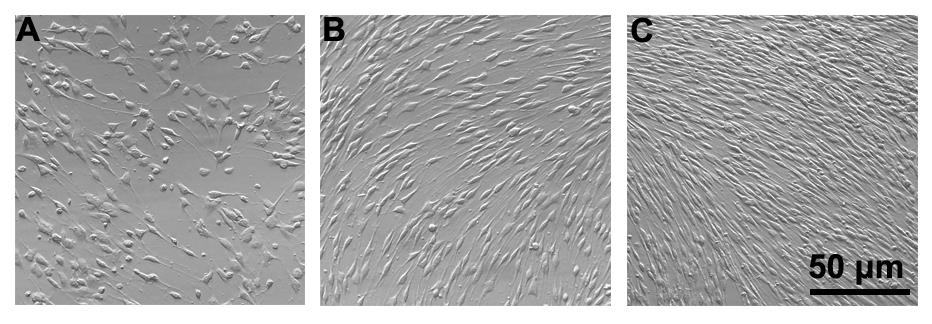
Figure 2. Rat Schwann cells’ purification and expansion. (A) Cells 24 h after dissection; the cultures should consist of Schwann cells and fibroblasts. Schwann cells have an elongated, spindle-shaped morphology, whereas fibroblasts are very flat. (B) On day 3, purified Schwann cells at confluence. (C) After being expanded in complete growth medium for two days and routinely passaged to the third passage in complete growth medium in the presence of forskolin and heregulin β-1, the culture is populated with Schwann cells (>95% confluence), with their characteristic bipolar, spindle-shaped cell bodies.
Differentiation and dedifferentiation of Schwann cells (Figure 3)
Seed and starvation: seed 5,000 Schwann cells in a 3.5 cm dish (covered with glass coverslips, for immunofluorescence analysis) or seed 1 × 10⁵ Schwann cells in a 6 cm dish (for western blotting analysis) in Medium A to allow a fast expansion of the cell population. After one day, remove the complete growth medium and rapidly replace it with an equal volume of Medium B overnight to adapt to the lack of mitogenic stimulation.
Note: Complete growth medium (Medium A) of Schwann cells containing forskolin will induce Schwann cells to acquire a differentiated phenotype, and 10% FBS will induce Schwann cells to proliferate rather than differentiate.
Differentiation: remove the starvation medium from each dish by gentle aspiration, replace it with an equal volume of Medium C and culture the cells in a CO2 incubator for 72 h. Observe the cultures of dbcAMP-differentiated Schwann cells under the phase contrast microscope to confirm the expected morphological changes prior to applying a dedifferentiating treatment.
Note: After approximately 6 h, you can start to see the morphological changes in rat Schwann cells. It is recommended to include cultures maintained without forskolin and dbcAMP from the outset to serve as controls. To maintain Schwann cells differentiated for an extended period of time, simply carry out a medium replacement using new differentiation conditional medium.
Dedifferentiation: after 72 h, the Schwann cells have differentiated in response to dbcAMP treatment. To induce dedifferentiation, simply replace the culture medium with Medium D, and culture the cells in a CO2 incubator for 24 h. Proceed to collect or analyze the cultures on the second day after dbcAMP withdrawal or as needed according to the experimental design.
Note: dbcAMP-induced differentiated Schwann cells cannot dedifferentiate when exposed to growth factors such as forskolin and heregulin β-1. Thus, it is recommended to use a medium that does not contain forskolin and heregulin β-1 throughout the differentiation and dedifferentiation assays. Approximately 6 h after dbcAMP removal, nearly half of rat Schwann cells return to a bipolar or tripolar shape. You may add a drug or perform virus transfection before the dedifferentiation assay.
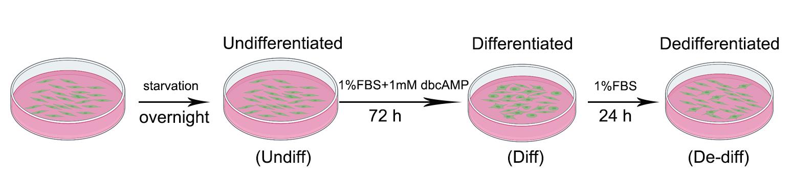
Figure 3. Differentiation and dedifferentiation system of rat Schwann cells. Schwann cells are expanded in Medium A for several days, after which the third-passage cells are replated at an appropriate density and starved overnight in Medium B. The next day, the cells are treated with 1 mM dbcAMP, which induces morphological change and upregulation of differentiation markers that can be examined by phase contrast microscope observation, immunofluorescence, or western blotting. After 72 h, simply replace the culture medium with Medium D to induce Schwann cell dedifferentiation. Undiff: Undifferentiated; Diff: Differentiated; De-diff: Dedifferentiated.
Data analysis
Phase contrast microscopy (Figure 4A)
To identify the phenotype of Schwann cells, observe their morphology under a phase contrast microscope. Schwann cells were treated with dbcAMP to obtain a differentiated phenotype with morphological transition from an elongated spindle-like shape to a flattened shape; simple dbcAMP removal can reverse the differentiated Schwann cells into an elongated bipolar morphology.
Immunofluorescence (Figure 4B)
Cell fixation: fix cells with 4% PFA for 30 min at room temperature.
Permeabilization and blocking: penetrate cells with 0.1% Triton X-100 (see Recipe 8) for 30 min and incubate with 5% gelatin (see Recipe 9) at room temperature for 1 h.
Primary antibodies: prepare a 1:200 dilution of mouse anti-c-Jun and a 1:100 solution of rabbit anti-Krox20 in 5% gelation solution. Incubate the cells with primary antibodies at 4 °C overnight.
Secondary antibodies: prepare a 1:400 dilution of Alexa Fluor® 488 goat anti-mouse IgG (H+L) or Alexa Fluor® 568 goat anti-rabbit IgG (H+L) in PBST (see Recipe 10) secondary antibodies. Incubate cells with the corresponding secondary antibodies at room temperature for 2 h, wash cells with PBST twice, and then incubate cells with 1 μg/mL DAPI for 5 min at room temperature to stain nuclei.
Western blotting (Figure 4C, 4D)
Cells lysis: add 100 μL of RIPA lysis buffer to the cell dish, blow, and collect lysates into a 1.5 mL tube.
Protein electrophoresis: prepare 10% dodecyl sulfate sodium salt-polyacrylamide gel, pipette 10 μg of protein into each well, and run the electrophoresis using the following parameters: 80 V and 300 mA for 1.5 h. When the electrophoresis is completed, remove the gel carefully and transfer proteins to a PVDF membrane.
Incubation of primary and secondary antibodies: after blocking with blocking buffer for 2 h at room temperature, prepare a 1:800 dilution of mouse anti-Sox2 and a 1:500 solution of rabbit anti-p75 in blocking buffer, incubate the membrane in primary antibody at 4 °C overnight, and incubate Horseradish peroxidase (HRP)-conjugated secondary antibody at room temperature for 2 h.
Visualization and calculation: visualize the membrane using Omni-ECLTM Light Chemiluminescence kit and calculate protein quantity using ImageJ.
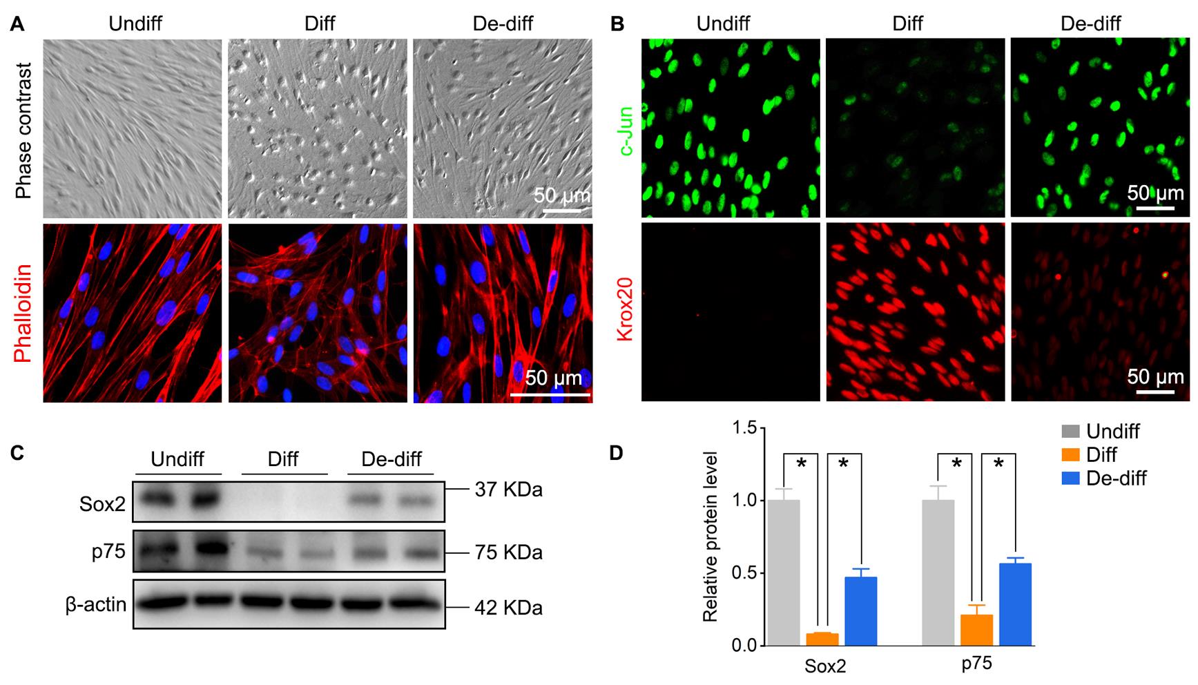
Figure 4. Identification of rat Schwann cells’ phenotype. (A) Cell morphology and cytoskeleton stained with phalloidin were observed under a microscope. (B) Immunofluorescence of c-Jun (indicates immature or dedifferentiated Schwann cells) and Krox20 (indicates mature or differentiated Schwann cells). (C, D) Western blotting analysis and data statistics of Sox2 and p75 (indicates immature or dedifferentiated Schwann cells). Undiff: Undifferentiated; Diff: Differentiated; De-diff: Dedifferentiated.
Recipes
1,000× Ara-C
Dissolve 4.86 mg of Ara-C in 2 mL of distilled water to make a 1,000× stock solution of 10 mM and sterilize the solution by filtration. Store at -20 °C.
1,000× dbcAMP
Dissolve 50 mg of dbcAMP in 101.756 μL of distilled DMSO to make a 1,000× stock solution of 1,000 mM and store at -20 °C.
30 mM forskolin stock solution
Dissolve 10 mg of forskolin in 812 μL of distilled DMSO to prepare a 30 mM forskolin stock solution. Store at -20 °C.
Complete growth medium of rat Schwann cells
48.5 mL of DMEM/F12 containing 1.5 mL of FBS, 3 μM forskolin, 10 ng/mL heregulin- β-1, and 100 mg/mL penicillin–streptomycin.
10% FBS
45 mL of DMEM/F12 containing 5 mL of FBS supplemented with 1% penicillin–streptomycin.
3% FBS
48.5 mL of DMEM/F12 containing 1.5 mL of FBS supplemented with 1% penicillin–streptomycin.
1% FBS
49.5 mL of DMEM/F12 containing 0.5 mL of FBS supplemented with 1% penicillin–streptomycin.
1 mM dbcAMP
Add 1 μL of 1,000× dbcAMP (1,000 mM) to 1 mL of 1% FBS.
0.1% Triton X-100
Dilute 1 mL of Triton X-100 into 1,000 mL of PBS.
5% gelatin
Dissolve 0.5 g of gelatin in 10 mL of PBS. Add 300 μL of Triton X-100 (0.3%) to the buffer.
PBST
Add 1 mL of Tween-20 to 1,000 mL of PBS. Store at room temperature.
Blocking buffer
5% non-fat milk in Tris-buffered saline containing 0.5% Tween-20
Acknowledgments
This protocol is adapted from the previous published papers (Zou et al., 2022) and (Wen et al., 2017). Thanks to Professor Jiasong Guo (Southern Medical University, Guangzhou, China).
Competing interests
There are no conflicts of interest or competing interests.
Ethics
All procedures involving animals were carried out with the approval of the Jinan University (Guangzhou, China) Animal Care and Use Committee in accordance with the guidelines for the ethical treatments of animals.
References
- Arthur-Farraj, P., Wanek, K., Hantke, J., Davis, C. M., Jayakar, A., Parkinson, D. B., Mirsky, R. and Jessen, K. R. (2011). Mouse schwann cells need both NRG1 and cyclic AMP to myelinate. Glia 59(5): 720-733.
- Cristobal, C. D. and Lee, H. K. (2022). Development of myelinating glia: An overview. Glia 70(12): 2237-2259.
- Jessen, K. R. and Mirsky, R. (2019). The Success and Failure of the Schwann Cell Response to Nerve Injury. Front Cell Neurosci 13: 33.
- Nocera, G. and Jacob, C. (2020). Mechanisms of Schwann cell plasticity involved in peripheral nerve repair after injury. Cell Mol Life Sci 77(20): 3977-3989.
- Wen, J., Tan, D., Li, L. and Guo, J. (2017). Isolation and Purification of Schwann Cells from Spinal Nerves of Neonatal Rat. Bio Protoc 7(20): e2588.
- Yamauchi, J., Miyamoto, Y., Hamasaki, H., Sanbe, A., Kusakawa, S., Nakamura, A., Tsumura, H., Maeda, M., Nemoto, N., Kawahara, K., Torii, T. and Tanoue, A. (2011). The atypical Guanine-nucleotide exchange factor, dock7, negatively regulates schwann cell differentiation and myelination. J Neurosci 31(35): 12579-12592.
- Zou, Y., Zhang, J., Liu, J., Xu, J., Fu, L., Ma, X., Xu, Y., Xu, S., Wang, X. and Guo, J. (2022). SIRT6 Negatively Regulates Schwann Cells Dedifferentiation via Targeting c-Jun During Wallerian Degeneration After Peripheral Nerve Injury. Mol Neurobiol 59(1): 429-444.
Article Information
Publication history
Accepted: Dec 25, 2022
Published: Mar 5, 2023
Copyright
© 2023 The Author(s); This is an open access article under the CC BY-NC license (https://creativecommons.org/licenses/by-nc/4.0/).
How to cite
Ying, Z. (2023). Establishment of an in vitro Differentiation and Dedifferentiation System of Rat Schwann Cells. Bio-protocol 13(5): e4631. DOI: 10.21769/BioProtoc.4631.
Category
Neuroscience > Peripheral nervous system > Schwann cell
Cell Biology > Cell isolation and culture > Cell differentiation
Cell Biology > Cell imaging > Fixed-cell imaging
Do you have any questions about this protocol?
Post your question to gather feedback from the community. We will also invite the authors of this article to respond.
![]() Tips for asking effective questions
Tips for asking effective questions
+ Description
Write a detailed description. Include all information that will help others answer your question including experimental processes, conditions, and relevant images.